May 1, 2026
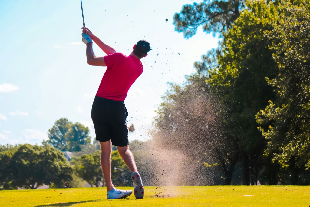
Golfer’s elbow is one of those injuries that often does not stop you playing straight away… It just annoyingly hangs around!It niggles on the range.It hurts when you grip the clubIt settles a bit, then flares again once you think you’re getting on top of it.That’s usually when golfers start trying random things — But...
Read more
November 28, 2025
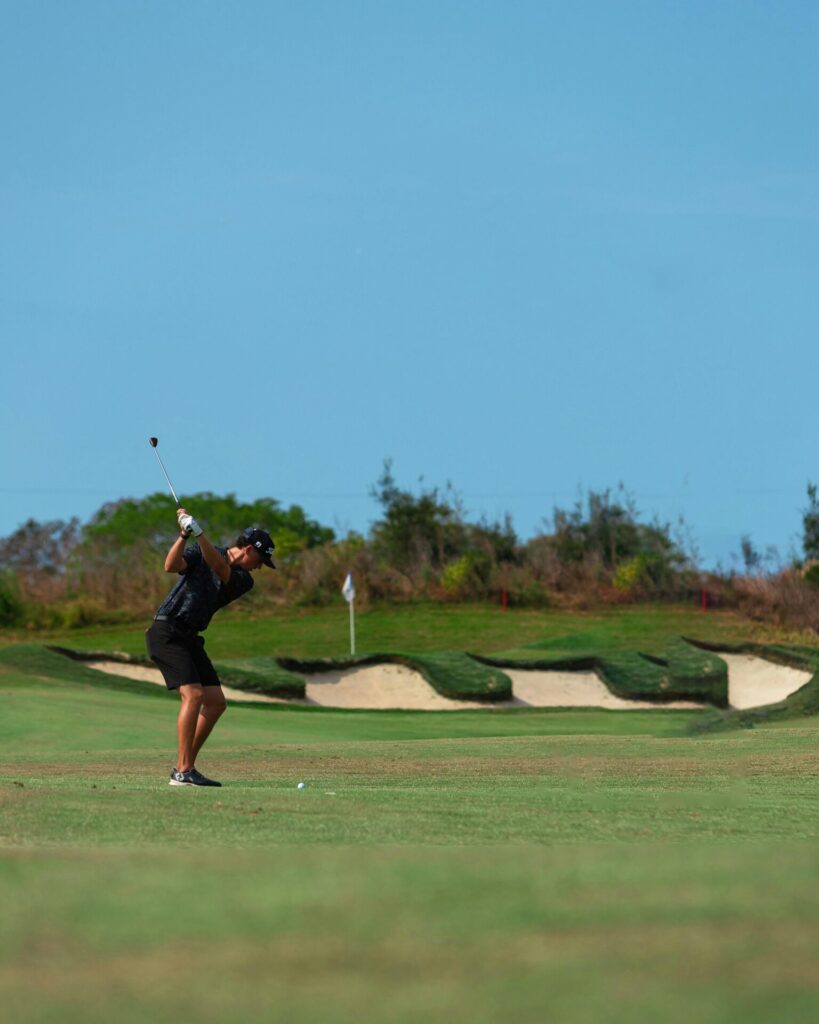
If you play golf around Caloundra or anywhere on the Sunshine Coast and feel like your body is holding you back, a golf physio screening can help answer whether it’s your swing or your body that needs attention.Maybe your back tightens up halfway through the front nine.Maybe your elbow nags for days after a bucket...
Read more
November 27, 2024
At Warana Sportscare, we’re proud to offer innovative treatments to help our patients heal and thrive. One such treatment is Shockwave Therapy. This cutting-edge therapy may sound complex, but it’s a simple and effective solution for a variety of conditions. What is Shockwave Therapy? Shockwave Therapy is short for Extracorporeal Shockwave Therapy (ESWT). In plain...
Read more
May 22, 2023
Pain leads to Panic, Panic leads to Fear, Fear leads to Suffering, Suffering leads to Pain. …and so the pain-cycle goes on and on! Years ago, I experienced what it was like to ride this pain-cycle while I battled through my own bout of persistent lower back pain. I remember what it was like,...
Read more
May 8, 2023
People in pain have generally done their research, talked to friends, and read everything they could find online. They form an expectation surrounding what their condition is and go on to develop a preferred treatment option. Now, this could be a good thing… or not. Patient expectations might not be accurate as they might not...
Read more
December 2, 2018
Follow Adam McKenzie our principal physiotherapist at Warana sportscare with his post operative recovery from a knee arthroscopy. Adam is now 10 days following a knee meniscus debridement and he will discuss his exercise progression and rehabilitation.
November 24, 2018
Follow our physiotherapist Adam McKenzie recover from his knee arthroscopy. Today he will discuss the progression of his exercise program for days 2 and 3 post surgery.
November 23, 2018
Follow Adam in his recovery and rehabilitation following a knee arthroscopy where he had a lateral meniscus debridement. This video is day 1 post op and Adam will keep you up to date of the exercises and recovery process.
August 16, 2017
Here’s part 2 in our series of yoga moves to help get the most out of every session in the surf. In this video Geoff focuses on warming up the spine.
July 25, 2017
Make sure you’re ready to surf your best as soon as you get in the water with our yoga activation exercises. Caloundra physiotherapist Geoff Ford has put together a series of activation exercises for surfers. Here is the first one.
December 11, 2014
Following a Medial Patello Femoral Ligament (MPFL) reconstruction or repair, the most important thing in the 48 hours after surgery is to try to get the swelling to settle down. You should rest and elevate the leg so your knee is above the level of your heart. This will help pain and swelling – don’t get up...
Read more
December 11, 2014
Exercises for the first two weeks after your ACL reconstruction In the first two weeks after your ACL reconstruction you should perform simple exercises aimed at boosting circulation and keeping muscles working during this time. In that first fortnight you should aim to get your knee out of its splint around three to four times. the...
Read more
December 11, 2014
Following your post-ACL reconstruction, you will experience some pain and swelling. The main idea in the first fortnight after your surgery is to get the pain and swelling under control. To do this, you need to rest with your leg elevated and use crutches at all times, not putting any weight on that leg. You...
Read more
December 11, 2014
Following a knee arthroscopy you should perform gentle exercises to get your knee moving. You can do these exercises laying on a flat surface. exercise one: tense thigh muscle and hold for five seconds exercise two: contract thigh muscles and lift leg exercise three: gentle knee bends exercise four: knee extensions with a rolled up towel under...
Read more
December 11, 2014
Recover quickly from your post-operative ankle arthroscopy To ensure a speedy and effective recovery from your ankle arthroscopy, your should rest, elevate and ice your ankle. Follow our tips below and watch our video. stack some pillows and elevate your ankle gently move your ankle up and down to boost circulation (flexes) try not to...
Read more
December 11, 2014
Following a knee arthroscopy, including microfracture, you need to remain non-weight bearing for six weeks and not put your foot to the ground at all during this time. You also shouldn’t bend your knee all the way to a 90-degree angle as this will put too much of a strain on the deep structures in...
Read more
October 11, 2014
Follow-up information following recent arthroscopy surgery When you come out of surgery you will have a compression bandage which goes above and below the knee. This bandage needs to be on for at least three to four days. The bandage is quite thick so there’s no point in icing the area while the bandage is...
Read more
June 11, 2014
Pelican Waters Swim Club core program Core exercises are important to help swimmers perform better and swim easier. This video demonstrates various levels of exercises to strengthen your core muscles. These exercises can be performed at home, all you need is a fit ball and a couple of balance disks and a straight stick which is...
Read more
January 11, 2014
Generic ankle strapping for a lateral ankle instability, i.e. if you have twisted your ankle. clean your skin to remove sweat and dirt start with an anchor point, not too tight, just a point to adhere more tape in different directions keep the foot flexed up then create some stirrups – always start from the...
Read more
December 11, 2009
Massage techniques parents can use on their swimmers at home These techniques are designed to improve specific movements swimmers need in order to swim fast. In behind the shoulder blade has a lot of trigger points and can get really tight. Get your swimmer to lay on their side and put their arm up and...
Read more